Living systems

Connected by nature. Distinct in biology.

Plants grow in living soil, and animals and people eat what grows. Inside each one, microbes work in a setting of their own, so each system is read with its own methods and its own evidence. The project follows humic and fulvic substances into each of them, because what a material does at a root is not what it does in a rumen or a colon, so each system is its own line of research.

The atlas

Six living systems, side by side.

Choose a system to see its scene, the microbial setting inside it, one useful question, and where its evidence and publications are.

  • Connected by nature. What really passes between systems: plants become forage, feed and food.

  • Distinct in biology. The same questions, asked separately in each host. The six are not stations on one line.

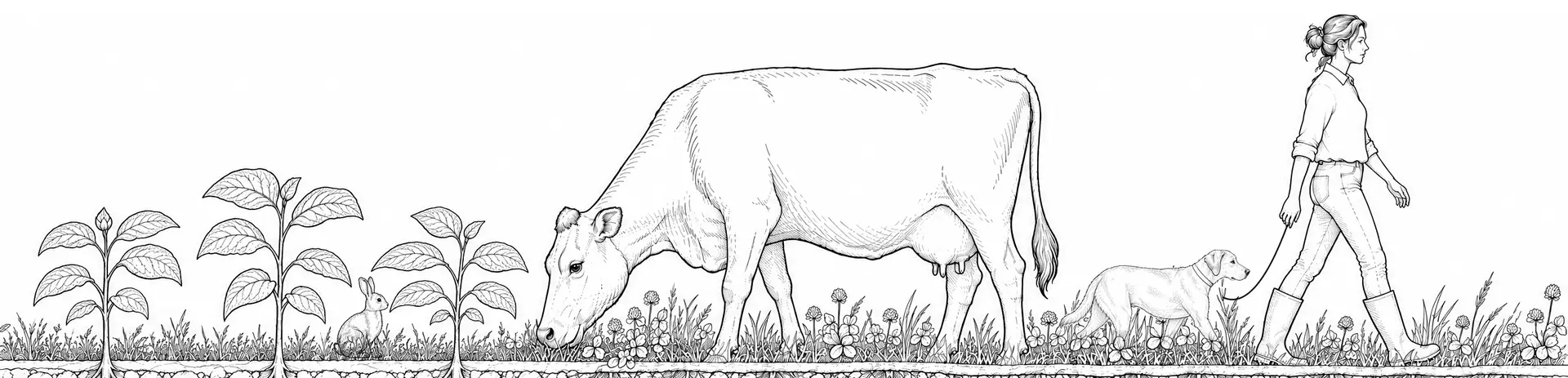
One field drawn in fine line: young plants with a rabbit, a cow grazing, and a woman walking a dog.
One field, drawn in fine line. Four of the six systems appear in it; the horse and the trout are chosen from the buttons below. Illustration, not to scale.

Soil & plants

The microbial setting
The rhizosphere: the soil closest to the root, where bacteria and fungi use the carbon compounds the root releases.
Connected by nature
What grows here becomes forage, feed and food for every other system on this page.
A useful question here
What changed in the root zone, and was it the soil, the season or the management?
Evidence
4 records in this collection. Open the soil and plants evidence
Publications
Guide, Plant and Soil Edition: coming soon. Handbook, Plant and Soil Edition: coming soon. See the publications
Explore soil and plants
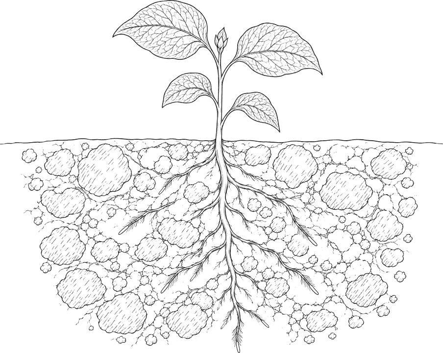
A young plant in outline above the soil line, its roots spreading through soil aggregates below.
Magnified view of the root zoneA root along the top with two root hairs reaching down into the soil, the carbon compounds the root releases (small dots), and bacteria clustered in the soil just below its surface. Fungal hyphae thread through the soil, one into a soil aggregate. A green tint marks the soil closest to the root, where microbial numbers and activity are highest.abcde

Magnified: the root zone

  • a Root
  • b Root exudates
  • c Bacteria
  • d Hyphae
  • e Aggregate

Above, the scene; below, a magnified view inside it. Illustration; magnified views are conceptual and not to scale.

Horses

The microbial setting
The hindgut: a large caecum and colon after the small intestine, where microbes ferment the forage the horse eats.
Connected by nature
A horse lives on forage: grass at pasture, or hay cut from it.
A useful question here
What changed in the horse’s forage, feeding and routine before the digestive change?
Evidence
One record in this collection. Open the horses evidence
Publications
Guide, Equine Edition: coming soon. Handbook, Equine Edition: coming soon. See the publications
Explore horses
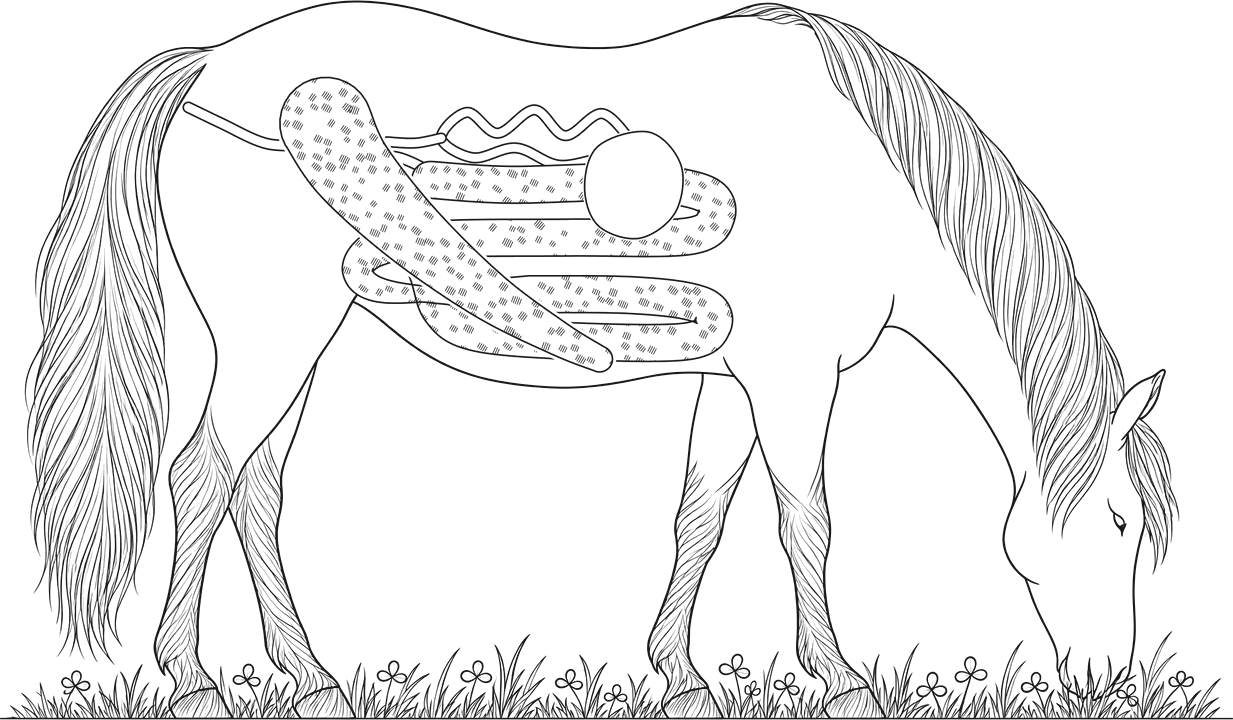
A grazing horse in outline with its digestive tract drawn inside.
Magnified view of the large colonThe wall of the large colon along the top, lined with mucus whose inner part holds few microbes. Below, fragments of forage fibre, drawn as rows of plant cell walls, with bacteria attached along their surfaces and gathered at their torn ends. A green tint marks the plant fibre that fibre-digesting microbes attach to and break down.abcd

Magnified: the large colon

  • a Large colon wall
  • b Plant fibre
  • c Bacteria
  • d Mucus layer

Above, the scene; below, a magnified view inside it. Illustration; magnified views are conceptual and not to scale.

Companion animals

The microbial setting
The large intestine. A dog eats a mixed diet; a cat is an obligate carnivore, so less plant fibre reaches its colon.
Connected by nature
Pet food is made from crops and from animals raised on farms.
A useful question here
Which change in food or routine came first, and in which animal?
Evidence
3 records in this collection. Open the companion animals evidence
Publications
Guide, Companion Animals: coming soon. Handbook, Companion Animals Edition: coming soon. See the publications
Explore companion animals
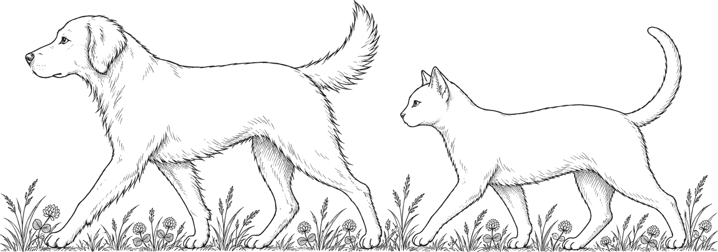
A dog and a cat walking in outline.
Magnified view of the large intestineThe colon wall along the top, lined with mucus whose inner part holds few microbes. Below, fragments of plant fibre and protein-rich residue from the diet, with bacteria attached along their surfaces and gathered at the torn ends of the fibre. A green tint marks the material that microbes attach to and break down.abcd

Magnified: the dog’s large intestine

  • a Colon wall
  • b Food residue and fibre
  • c Bacteria
  • d Mucus layer

Above, the scene; below, a magnified view inside it. Illustration; magnified views are conceptual and not to scale.

Livestock

The microbial setting
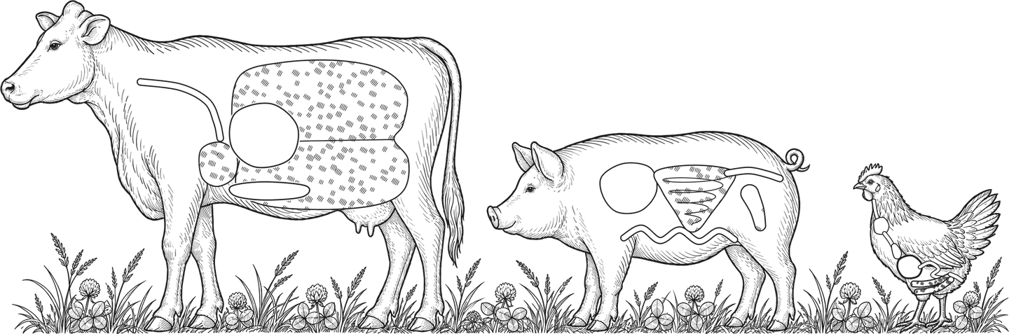
Three fermentation chambers: the cow’s rumen before the true stomach, the pig’s hindgut, and the hen’s two caeca.
Connected by nature
Cattle graze and eat forage; pigs and poultry eat feed grown as crops.
A useful question here
Where does this animal ferment its feed, and was that where the measurement was taken?
Evidence
5 records in this collection. Open the livestock evidence
Publications
Guide, Dairy Operations Edition: coming soon. 5 handbooks, by operation, coming soon. See the publications
Explore livestock
A cow, a pig and a hen in outline with their main fermentation chambers drawn inside.
Magnified view of the rumenThe upper wall of the rumen along the top, with a few short papillae and bacteria attached to its surface. Below, fragments of plant fibre, drawn as rows of plant cell walls, with bacteria attached along their surfaces and gathered at their torn ends, and two protozoa in the fluid between. A green tint marks the plant fibre that fibre-digesting microbes attach to and break down.abcd

Magnified: the cow’s rumen

  • a Rumen wall
  • b Plant fibre
  • c Bacteria
  • d Protozoa

Above, the scene; below, a magnified view inside it. Illustration; magnified views are conceptual and not to scale.

Aquaculture

The microbial setting
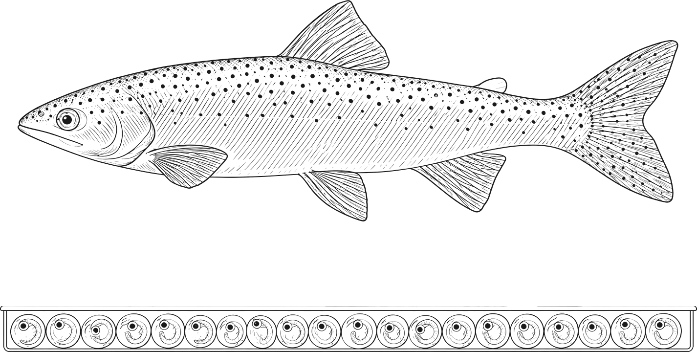
The trout intestine, with folds rather than villi and a sparser community than a mammal’s hindgut, and the water around the fish and its eggs.
Connected by nature
Feed for farmed trout is made in part from crops, and the fish lives in managed water.
A useful question here
Was the change measured in the water, the tank or the fish?
Evidence
2 records in this collection. Open the aquaculture evidence
Publications
Guide, Aquaculture Operations Edition: coming soon. Handbook, Aquaculture Operations Edition: coming soon. See the publications
Explore aquaculture
A rainbow trout in outline above a row of eggs in a tray.
Magnified view of the trout intestineThe intestinal wall along the top with its mucosal folds, each lined with mucus. Below, feed particles with bacteria attached to their surfaces; the community is sparser than in a mammal's hindgut. A green tint marks the feed particles bacteria attach to; the trout digests them mainly with its own enzymes.abcd

Magnified: the trout intestine

  • a Gut wall and folds
  • b Feed particle
  • c Bacteria
  • d Mucus layer

Above, the scene; below, a magnified view inside it. Illustration; magnified views are conceptual and not to scale.

Humans

The microbial setting
The large intestine, where bacteria ferment the dietary fibre that reaches it.
Connected by nature
People eat what grows in soil and what is raised on farms.
A useful question here
Does the study’s group, preparation and measure match the question being asked?
Evidence
3 records in this collection. Open the humans evidence
Publications
No edition for this system yet. Read what is available
Explore humans
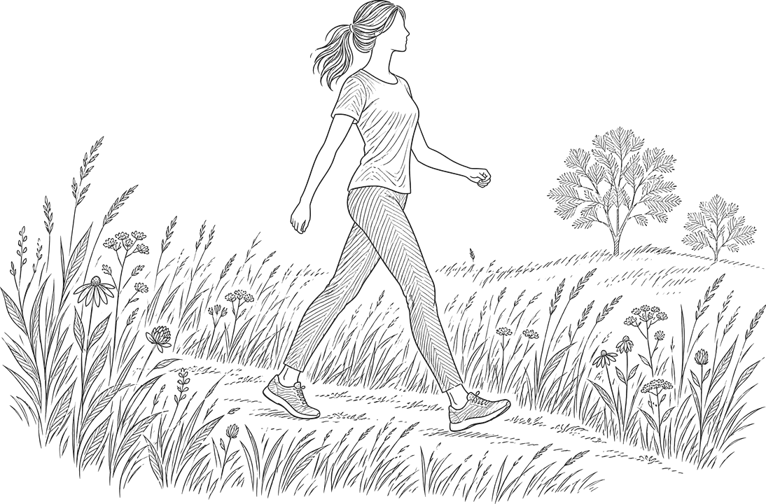
A woman walking along a meadow path.
Magnified view of the large intestineThe colon wall along the top, lined with mucus whose inner part holds few microbes. Below, fragments of plant fibre, drawn as rows of plant cell walls, with bacteria attached along their surfaces and gathered at their torn ends. A green tint marks the plant fibre that fibre-digesting microbes attach to and break down.abcd

Magnified: the large intestine

  • a Colon wall
  • b Plant fibre
  • c Bacteria
  • d Mucus layer

Above, the scene; below, a magnified view inside it. Illustration; magnified views are conceptual and not to scale.

All six systems as a list
  • Soil & plants

    Microbial setting. The rhizosphere: the soil closest to the root, where bacteria and fungi use the carbon compounds the root releases.

    Connected by nature. What grows here becomes forage, feed and food for every other system on this page.

    A useful question. What changed in the root zone, and was it the soil, the season or the management?

    Evidence. 4 records in this collection. Open the evidence

    Publications. Guide, Plant and Soil Edition: coming soon. Handbook, Plant and Soil Edition: coming soon. See the publications

  • Horses

    Microbial setting. The hindgut: a large caecum and colon after the small intestine, where microbes ferment the forage the horse eats.

    Connected by nature. A horse lives on forage: grass at pasture, or hay cut from it.

    A useful question. What changed in the horse’s forage, feeding and routine before the digestive change?

    Evidence. One record in this collection. Open the evidence

    Publications. Guide, Equine Edition: coming soon. Handbook, Equine Edition: coming soon. See the publications

  • Companion animals

    Microbial setting. The large intestine. A dog eats a mixed diet; a cat is an obligate carnivore, so less plant fibre reaches its colon.

    Connected by nature. Pet food is made from crops and from animals raised on farms.

    A useful question. Which change in food or routine came first, and in which animal?

    Evidence. 3 records in this collection. Open the evidence

    Publications. Guide, Companion Animals: coming soon. Handbook, Companion Animals Edition: coming soon. See the publications

  • Livestock

    Microbial setting. Three fermentation chambers: the cow’s rumen before the true stomach, the pig’s hindgut, and the hen’s two caeca.

    Connected by nature. Cattle graze and eat forage; pigs and poultry eat feed grown as crops.

    A useful question. Where does this animal ferment its feed, and was that where the measurement was taken?

    Evidence. 5 records in this collection. Open the evidence

    Publications. Guide, Dairy Operations Edition: coming soon. 5 handbooks, by operation, coming soon. See the publications

  • Aquaculture

    Microbial setting. The trout intestine, with folds rather than villi and a sparser community than a mammal’s hindgut, and the water around the fish and its eggs.

    Connected by nature. Feed for farmed trout is made in part from crops, and the fish lives in managed water.

    A useful question. Was the change measured in the water, the tank or the fish?

    Evidence. 2 records in this collection. Open the evidence

    Publications. Guide, Aquaculture Operations Edition: coming soon. Handbook, Aquaculture Operations Edition: coming soon. See the publications

  • Humans

    Microbial setting. The large intestine, where bacteria ferment the dietary fibre that reaches it.

    Connected by nature. People eat what grows in soil and what is raised on farms.

    A useful question. Does the study’s group, preparation and measure match the question being asked?

    Evidence. 3 records in this collection. Open the evidence

    Publications. No edition for this system yet. Read what is available

Shared questions

What conditions are present? What are microbes doing? What can we observe?

The questions are the same in every system. The methods are not.

  • Root zones

    Microbes work at the root surface, in soil that changes over a short distance and across a season. Methods sample roots and soil and record the site, the weather and the management.

  • Rumens

    Microbes ferment feed before it reaches the true stomach. Methods sample the rumen fluid itself and read it against the ration.

  • Hindguts

    In horses and pigs, fermentation happens after the small intestine. Faeces are easy to collect, but they reflect the hindgut only in part.

  • Water systems

    For fish, the water is part of the setting. A dose is stated per litre, and a change can be measured in the water, the tank or the animal.

  • Human intestines

    The large intestine is studied through diet records, stool samples and ethically reviewed trials. A finding applies to the people who took part.

Evidence from one setting can suggest a question in another. It does not answer it.

Bring a field-specific perspective.

The project is developing through education, evidence review and opportunities for collaboration. We welcome expertise that helps make the questions more precise and the interpretations more useful.

The waiting list

Join the waiting list

Leave your email and we will send it to you when it is ready.

One email when your edition is ready. Nothing else.